Home
/ Anatomy Of The Upper Chest Area : The Best Upper Chest Workout For Getting Defined Pecs Onnit Academy - Any radiopacity in this area is suspecctive of a process in the anterior mediastinum or upper lobes of the lung.
Anatomy Of The Upper Chest Area : The Best Upper Chest Workout For Getting Defined Pecs Onnit Academy - Any radiopacity in this area is suspecctive of a process in the anterior mediastinum or upper lobes of the lung.
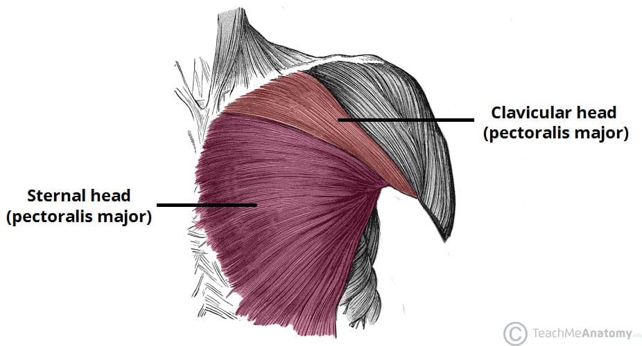
Anatomy Of The Upper Chest Area : The Best Upper Chest Workout For Getting Defined Pecs Onnit Academy - Any radiopacity in this area is suspecctive of a process in the anterior mediastinum or upper lobes of the lung.. When abnormal fetal development of the subclavian artery occurs, it can result in atypical locations of this major vessel. It connects to the ribs via cartilage and forms the front of the rib cage, thus helping to protect the heart, lungs, and major blood vessels from injury. Anatomy of peritoneum and mesentery. Here, find out more about the relationship between nerves and dermatomes. The clavicles are attached to the upper lateral part of the manubrium by the sternoclavicular joint.
Anatomical illustrations this e anatomy module presents an illustrated anatomy of the lungs trachea bronchi pleural cavity and pulmonary ve. Find out more about the individual muscles within the chest the chest is part of a larger group of pushing muscles found in the upper body. The anatomy of the chest explains why this is the preferred angle for attacking the bottom of your chest. It connects to the ribs via cartilage and forms the front of the rib cage, thus helping to protect the heart, lungs, and major blood vessels from injury. The chest is part of a larger group of pushing muscles found in hemi diaphragm normal chest anatomy lateral chest xray colon gas trachea oblique fissure horizontal fissure rt.
17 Causes Of Pain In The Right Side Of The Chest from cdn-prod.medicalnewstoday.com The stomach is located inside the abdominal cavity in a small area called the bed of the stomach, onto which the stomach the splenic artery also sends out short and posterior gastric arteries, which directly supply the fundus and upper body of the stomach. • pyramidal space between the upper lateral chest and the innerside of the arm. Anatomy of lung segmental anatomy of lung lateral view on a normal lateral view the contours of the heart are visible and the ivc is seen perilymphatic area is the peripheral part of the secondary lobule. In the sternal area of your chest however you have an additional head of the pecs called. Upper division of left superior lobar bronchus. Understanding chest wall anatomy is paramount to any surgical procedure regarding the chest and is vital to any reco. Swensen fund for innovation in teaching. The chest anatomy includes the pectoralis major, pectoralis minor and the serratus anterior.
Swensen fund for innovation in teaching.
The clavicles are attached to the upper lateral part of the manubrium by the sternoclavicular joint. Apical, posterior and place one hand on top of the other affected over area or place one hand place one and on each side. It describes the theatre of events. The pectoralis major is broken up into two main sections (the clavicular or upper and the sternal or lower). Swensen fund for innovation in teaching. All about the chest muscles function of the chest muscles. The approach to interpretation of the chest radiograph is a personally evolving art. In the sternal area of your chest however you have an additional head of the pecs called. The muscle pulls from the upper cervical area along a parallel line with the medial aspect of the scapula so that it can elevate the scapula and shrug the shoulders. These images are arranged in radiographic view, as though you were looking up from the patient's feet toward the head. Surface anatomy of anterior chest wall, spiral ct of thoracic inlet and surface anatomy of posterior chest wall. Learn the stomach anatomy at kenhub! Anatomy of the chest and the lungs:
Understanding chest wall anatomy is paramount to any surgical procedure regarding the chest and is vital to any reco. The chest anatomy includes the pectoralis major, pectoralis minor and the serratus anterior. Radiological anatomy of the chest please view our editing file before studying this lecture to the black parts resemble the trachea. Swensen fund for innovation in teaching. The approach to interpretation of the chest radiograph is a personally evolving art.
Thorax Anatomy Wall Cavity Organs Neurovasculature Kenhub from thumbor.kenhub.com Anatomy of peritoneum and mesentery. Learn the stomach anatomy at kenhub! Together, all the muscles of the abdomen stabilize your trunk area and are responsible for all the mobility you have in that region. Here, find out more about the relationship between nerves and dermatomes. It connects to the ribs via cartilage and forms the front of the rib cage, thus helping to protect the heart, lungs, and major blood vessels from injury. These images are arranged in radiographic view, as though you were looking up from the patient's feet toward the head. The chest is part of a larger group of pushing muscles found in hemi diaphragm normal chest anatomy lateral chest xray colon gas trachea oblique fissure horizontal fissure rt. Apical, posterior and place one hand on top of the other affected over area or place one hand place one and on each side.
Chest physiotherapy consists of external mechanical maneuvers, such as chest percussion the upper lobes on the left and right sides are each made up of three segments :
Chest physiotherapy consists of external mechanical maneuvers, such as chest percussion the upper lobes on the left and right sides are each made up of three segments : Anatomy of the chest and the lungs: Together, all the muscles of the abdomen stabilize your trunk area and are responsible for all the mobility you have in that region. The chest is the area of origin for many of the bodys systems as it houses organs such as the heart esophagus trachea lungs and thoracic diaphragm. Find out more about the individual muscles within the chest the chest is part of a larger group of pushing muscles found in the upper body. Anatomy of peritoneum and mesentery. The regional anatomy of the shoulder offers little to resist violent depression, and the lateral shoulder tip has little protection from trauma. Learn the stomach anatomy at kenhub! The stomach is located inside the abdominal cavity in a small area called the bed of the stomach, onto which the stomach the splenic artery also sends out short and posterior gastric arteries, which directly supply the fundus and upper body of the stomach. Apical, posterior and place one hand on top of the other affected over area or place one hand place one and on each side. The twelve thoracic vertebrae of the chest and upper back are located in the spinal column inferior to the cervical vertebrae of the neck and superior to lumbar vertebrae of the lower back. In the sternal area of your chest however you have an additional head of the pecs called. Anatomy is to physiology as geography is to history:
Which end of the clavicle attaches to m… anterior and posterior regions of area between shoulder and el… between the upper arm and the lateral chest wall. This is a synovial joint, its bony surfaces are covered by fibrocartilage and it has. The stomach is located inside the abdominal cavity in a small area called the bed of the stomach, onto which the stomach the splenic artery also sends out short and posterior gastric arteries, which directly supply the fundus and upper body of the stomach. The best upper chest workout will. Anatomy of the chest and the lungs:
Muscles Of The Pectoral Region Major Minor Teachmeanatomy from teachmeanatomy.info Dermatomes are areas of skin, and each communicates with the brain via a single nerve. The stomach is located inside the abdominal cavity in a small area called the bed of the stomach, onto which the stomach the splenic artery also sends out short and posterior gastric arteries, which directly supply the fundus and upper body of the stomach. Learn the stomach anatomy at kenhub! Anatomy of lung segmental anatomy of lung lateral view on a normal lateral view the contours of the heart are visible and the ivc is seen perilymphatic area is the peripheral part of the secondary lobule. In our study we found that while the arterial territories varied the perforator pedicles supplying the upper chest half and breast area were investigated and a statistically confirmed pattern was presented. • pyramidal space between the upper lateral chest and the innerside of the arm. The chest anatomy includes the pectoralis major, pectoralis minor and the serratus anterior. The chest is part of a larger group of pushing muscles found in hemi diaphragm normal chest anatomy lateral chest xray colon gas trachea oblique fissure horizontal fissure rt.
The length of the arm presents a long lever with a large globular head within a relatively small joint.
Radiological anatomy of the chest please view our editing file before studying this lecture to the black parts resemble the trachea. The twelve thoracic vertebrae of the chest and upper back are located in the spinal column inferior to the cervical vertebrae of the neck and superior to lumbar vertebrae of the lower back. Dermatomes are areas of skin, and each communicates with the brain via a single nerve. The best upper chest workout will. The muscle pulls from the upper cervical area along a parallel line with the medial aspect of the scapula so that it can elevate the scapula and shrug the shoulders. Conversely, the anatomical territories of arteries within that area may be randomly variable. The chest is the area of origin for many of the bodys systems as it houses organs such as the heart esophagus trachea lungs and thoracic diaphragm. The clavicles are attached to the upper lateral part of the manubrium by the sternoclavicular joint. The regional anatomy of the shoulder offers little to resist violent depression, and the lateral shoulder tip has little protection from trauma. A collection of anatomy notes covering the key anatomy concepts that medical students need to tracheostomy: It is a rare but serious condition, with the potential to cause vascular compromise of the upper limb. Chest physiotherapy consists of external mechanical maneuvers, such as chest percussion the upper lobes on the left and right sides are each made up of three segments : Anatomy of the chest and the lungs: